Medical Research Institute KITANO HOSPITAL, PIIF Tazuke-Kofukai
Inquiry
search
close

Departments
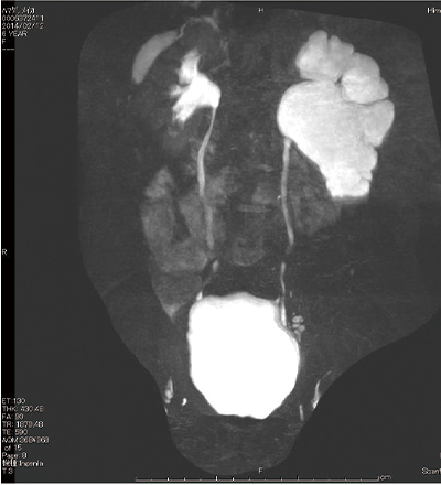
Urine is produced in the kidneys, collected in a chamber called the renal pelvis, and then travels through the ureters to the bladder for storage. This condition occurs when the area where the renal pelvis and ureter meet is narrow, causing obstruction to the bladder. When pressure within the renal pelvis increases and the pelvis expands, this condition is called hydronephrosis. Recently, it has often been detected during ultrasound scans during pregnancy. Most cases are asymptomatic and resolve spontaneously. However, there are some cases where the condition does not resolve spontaneously and leads to a decline in kidney function, in which case surgical treatment is required.
Depending on the degree of hydronephrosis (dilation of the renal pelvis), it is classified into grades I to IV.
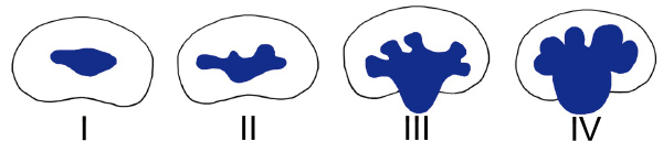
Grade of hydronephrosis
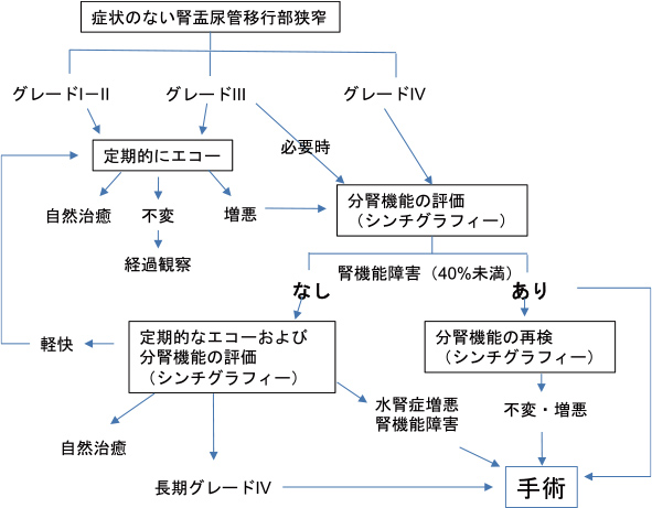
*From the 2016 Clinical Practice Guide for Pediatric Congenital Hydronephrosis (Ureteropelvic Junction Obstruction)
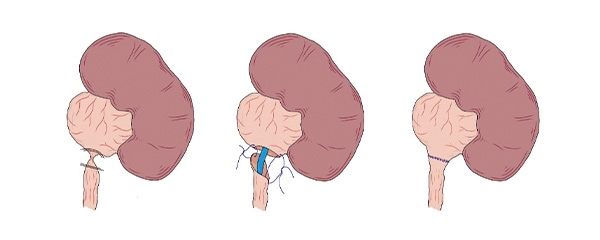
Depending on the patient's physique, surgery can be performed using open surgery, laparoscopic surgery, or robotic-assisted surgery. During surgery, a tube called a stent is placed between the renal pelvis and the bladder. The stent is removed using a cystoscope around two months after surgery.
Regular checkups and tests will be conducted after surgery. It takes time for hydronephrosis to improve after surgery.