Medical Research Institute KITANO HOSPITAL, PIIF Tazuke-Kofukai
Inquiry
search
close

Departments
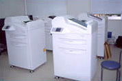
All imaging is done digitally using a CR (manufactured by FUJI), with the exception of the Health Management Center, which uses flat panel digital imaging (manufactured by Canon).
CR is an abbreviation for Computed Radiography, which in Japanese is called "computer X-ray photography." Nowadays, the mainstream CR system uses an imaging plate instead of a film coated with photosensitive material.
Digital images taken with CR can be read directly on a monitor, or they can be output onto film using a dry imager to produce what is commonly known as an X-ray, which can then be read on a scanning screen.
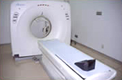
CT is an abbreviation for Computed Tomography, which means "computerized tomography" in Japanese. The donut-shaped section of the scanner typically contains a pair of X-ray generating tubes and a detector that measures the amount of X-rays emitted from the tubes and passed through the body, positioned facing each other. The X-rays continuously emitted from the tube are captured by the detector on the opposite side as the scanner rotates around the patient's body, and cross-sectional images of the body's internal structures are constructed by a computer, allowing for a detailed examination of the body's internal structures.
Recently, by increasing the number of detectors and taking continuous spiral scans in the length direction (helical scan), it has become possible to process large amounts of X-ray data quickly at once, making it possible to construct not only cross-sectional images but also three-dimensional images.
Our hospital uses two 64-channel multi-detector CT machines (manufactured by Toshiba) that enable high-speed scanning and the creation of highly accurate 3D images.
In addition, a 16-channel MDCT (manufactured by Yokogawa GE) is used for treatment planning.
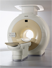
MRI is an abbreviation for Magnetic Resonance Imaging, which is called "nuclear magnetic resonance imaging" in Japanese. It is a device that applies RF pulses (radio waves used in radios, etc.) to the human body in a very strong magnetic field, and creates images by utilizing the phenomenon that different tissues return different signals. Therefore, unlike CT scans which use X-rays, MRI does not involve radiation exposure.
Furthermore, by using an imaging program (a process that converts signal values into an image), it is possible to obtain images that correspond to various information about the organ being imaged and the expected state of the lesion.
Currently, Philips uses three 1.5 Tesla MRI machines and one 3 Tesla machine. Compared to the 1.5 Tesla, the 3 Tesla machine can take clearer images, but it is susceptible to the effects of body movement, dielectric effects (interference between the radio waves that first pass and the reflected radio waves), and changes in magnetic susceptibility at the boundaries of metal and tissue, so in some cases the 1.5 Tesla machine may produce better image quality.
While the 3 Tesla is primarily used for examinations of the head, joints, blood vessels, and breasts, we also use both devices to examine all body parts, taking into consideration the purpose of the examination and the patient's condition.
*Tesla: A unit of magnetic field strength. Generally, the larger the unit, the higher the contrast/noise ratio and the better the image.
We use four DR devices (manufactured by Hitachi), all of which are digital and require minimal radiation exposure. We also use a high-resolution multipurpose fluoroscopy table.
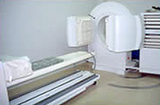
This is an examination in which radiation emitted from the body when a radioactive isotope (RI) agent that emits radiation is captured by a device called a scintigraphy camera, and the image is processed by computer to create an image.
Also known as an RI test, scintigram, nuclear medicine test, or isotope test, this test examines the strength, temporal changes, and distribution of radiation within the body to learn about changes in the shape and movement of each organ.
By observing the distribution of RI, detailed information can be obtained about abnormalities in the thyroid gland, lungs, liver, adrenal glands, etc., the condition of bones throughout the body, and the state of blood flow in the brain and myocardium.
Our hospital uses two RI machines: one with two heads (manufactured by Hitachi-Adac) and one with two heads (manufactured by Hitachi-Philips).
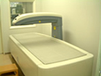
We conduct examinations using the most reliable DEXA-type X-ray bone density measuring device (PRODIGY).
This device is highly accurate and is used to diagnose osteoporosis and determine the effectiveness of treatment.