Kitano Medical Research Institute, Public Interest Incorporated Foundation, Tatsunokofukai
Inquiry
search
close
Have you ever had blue lumps on your calves or felt fatigue in your legs?
Human blood vessels consist of arteries that travel from the heart to the periphery and veins that return from the periphery to the heart. In the veins of the lower limbs, blood returns to the heart against gravity, thanks to the pumping action of the calf muscles and the valves in the veins. Varicose veins occur when these valves are damaged, allowing venous blood to flow backward, causing the veins to expand abnormally and become tortuous.
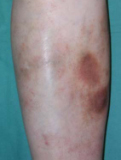
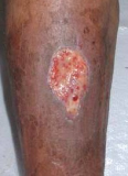
Natural history of varicose veins
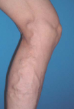
Left: Typical varicose veins
Medium: Pigmentation
Right: Intractable skin ulcer
It is often seen in people who work standing up for long periods of time or who suffer from illnesses such as high blood pressure or heart failure. Some women develop the condition during pregnancy and experience gradual worsening thereafter. It typically manifests as a soft, bluish bump on the inside of the calf. Prolonged standing or walking can cause the legs to feel fatigued and require rest (intermittent claudication). As the condition worsens, dull brown pigmentation appears, and over time, sores and wounds (skin ulcers) can develop. These skin ulcers are extremely painful and do not improve easily even with ointment treatments, making them known as intractable skin ulcers. Surgery is strongly recommended for those with pigmentation or skin ulcers.
An ultrasound (echo) examination is used to check whether the venous valves that prevent blood from flowing backward are functioning properly. This examination is painless, as it simply involves applying a jelly and placing a probe called a probe. We also check the diameter of the veins and the time it takes for the blood to flow backward, which are used as indicators for selecting a treatment method. We can also check whether you have had deep vein thrombosis in the past or present. Angiography using tomography may also be performed to understand the overall condition of the blood vessels. This involves taking an MRI while an intravenous contrast agent is administered.
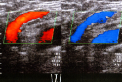
An ultrasound examination in action (red indicates reflux of blood)
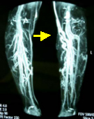
Actual MRI angiography (arrow indicates varicose veins)
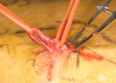
Partial vein removal surgery
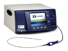
High-frequency treatment equipment for intravascular cauterization